In ultrasound diagnostics, there is a clear demand for quick and accurate diagnosis the first time and in less time. Philips EPIQ ultrasound platform with PercuNav allows clinicians to fuse CT/MR/PET images with live ultrasound so they can make confident decisions even in challenging diagnostic cases. In this case study, Prof. Dirk-André Clevert, MD, Associate Professor of Radiology at Munich University Hospital Grosshadern, tells how PercuNav used with contrast-enhanced ultrasound (CEUS) supports more confident diagnoses and can even change the course of treatment. This material is not for distribution in the USA
The clinical need
CT and MRI exams do not always provide enough information for a confident diagnosis or to determine the potential progression of lesions. In addition, these exams and the contrast agents used with them can be expensive. CT and MR contrast agents are also potentially nephrotoxic, which can be of concern in an older patient population. In comparison, contrast-enhanced ultrasound is a very inexpensive examination and without nephrotoxity risks, although visualizing inconspicuous complex lesions under contrast-enhanced ultrasound alone can also be technically challenging. Download the case study from Prof. Clevert.
When you combine the advantages of all the other modalities with ultrasound, at the end of the day it makes a better diagnosis for the patient.”
Dirk-André Clevert
MD

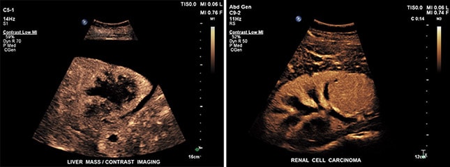
Contrast image of liver mass (left) and renal cell carcinoma (right)
Adding value to existing images
Fully integrated fusion capabilities mean that Prof. Clevert can now use multimodality query retrieve to view DICOM images such as CT and MR, allowing him to easily compare past and current studies without the use of an external reading station and even review these multimodality images while doing live imaging.

The image above shows a fusion image of a liver cyst (left) and a fusion image of a pancreas (right) using PercuNav. Images courtesy of Dr. Clevert.
For unclear lesions we rule out what kind of lesion it might be. Even when we perform high-end CT or MR scans there are cases in which the lesions are unclear. In addition, outpatient CT exams can sometimes be of poorer quality, at times due to patient movement or scanning limitations. We don’t repeat the CT or MR examination, instead we make use of the existing CT or MR image with additional fusion.”
Dirk-André Clevert
MD
Benefits of fusion imaging using ultrasound


