
UBC researchers advance their multiple sclerosis imaging
FieldStrength MRI magazine
User experiences - November 2019
Switching to Ingenia Elition has created new opportunities for MS imaging at the University of British Columbia
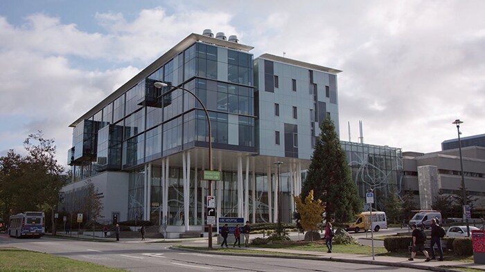
One of the main research areas at the MRI Research Centre at the University of British Columbia (UBC), in Vancouver Canada, is imaging of multiple sclerosis (MS). The center runs many studies and trials, and its researchers continually strive to improve and expand imaging methods to help in visualizing the disease and support diagnosis and monitoring. In this article, UBC researchers explain how their recently installed 3.0T MRI system, a Philips Ingenia Elition, helps them advance their MSrelated research studies. At the same time, they feel this scanner makes their lives easier and surprises them with the impact it has on patients and volunteers.
“Switching from our old scanner to Elition is like jumping from a flip phone to the latest smart phone”
Alex MacKay, DPhil
Professor of Physics, with a joint appointment in the Department of Radiology and the Department of Physics and Astronomy at the University of British Columbia, and Director of the UBC MRI Research Center. He has been working with MRI since about 1988 with a main research focus on neuro.
Alex Rauscher, PhD
Associate Professor and physicist at the University of British Columbia. His research focus is on quantitative brain MRI and includes mapping tissue susceptibility, myelin mapping, and studying the effects of anisotropic tissue orientation on MRI.
Shannon Kolind, PhD
Assistant Professor and physicist at the University of British Columbia, currently working in the Department of Medicine, division of neurology. Her main goal is translating current quantitative- and biologicallyspecific MRI methods for use in clinical environment, in particular myelin imaging for MS.
Laura Barlow, RTMR
MRI Technologist Supervisor at the University of British Columbia MRI Research Center, 3T facilities. She has been working with MRI for a decade. Her current role focuses on the field of advanced neuro research.
Trends in visualizing and identifying MS lesions with MRI
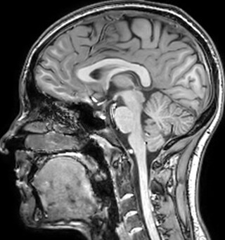
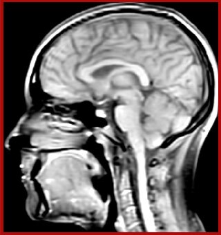
Multiple sclerosis or MS is a neurodegenerative disease that is characterized by myelin degradation, resulting in cognitive and motor deficits. According to Dr. Shannon Kolind, MR imaging for diagnosis and monitoring of MS is moving to higher field strength and using more 3D sequences, as reflected in the CMSC guidelines [1-3].
At the UBC MRI Research Centre, which is currently staffed by 70 principal investigators, the majority of studies and clinical trials are brain research, with MSrelated research as a principle area of interest. The center installed a 3.0T Ingenia Elition scanner in December 2018 as a successor of their Achieva 3.0T, with the intention to use it for all future research studies.
“In addition to traditional imaging like FLAIR for lesion identification, we see a real push towards techniques that weren’t normally required for MS, including good highresolution 3D T1 weighted images to do volumetrics. We’ve also started looking at spinal cord imaging again, since techniques have improved in terms of acquisition and analysis. Another important technique is susceptibility weighted imaging (SWI), particularly if we are looking for central veins in lesions, which is extremely helpful for diagnosis.”
Boosting myelin imaging capabilities to help visualize MS progression
At UBC, a lot of the MS-related work focuses on myelin imaging. “We're born with very little myelin and that increases as the brain grows, which is important for nerve signal propagation. Multiple sclerosis on the other hand, degenerates the myelin with the opposite effect. So, myelin has a really important role in brain function, and having a tool that measures myelin can be extremely useful, we feel,” says Dr. MacKay.
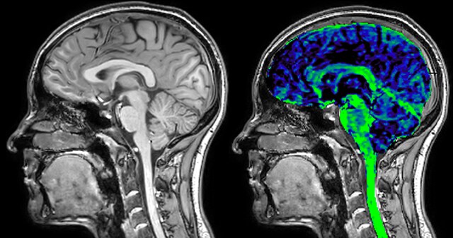
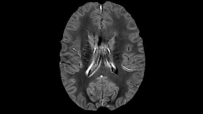
Myelin water imaging (MWI) is a breakthrough technique that was pioneered at UBC for measuring myelin content in the brain, in vivo. “Because the T2 time of water in myelin is much shorter than the T2 of water in the intraand extracellular spaces, we can separate out the myelin water signal.”
The techniques for measuring myelin have changed a lot over the years. “Since we are using the Elition, our myelin water images are much better. We're now acquiring 1 x 2 x 5 mm voxels and displaying at 1 x 1 x 2.5 mm. For a whole brain we can now measure the fraction of water in the myelin component in only about five or six minutes,” Dr. MacKay says.
“We are actually starting to apply this technique in clinical trials,” says Dr. Kolind. “And I’m really hopeful that this is going to give us more clues about the underlying disability progression in MS and move us towards some treatment options for that aspect of the disease.”
Dr. MacKay adds that researchers at UBC are also involved in trials to examine the effects of MS drug treatments on myelination. “In a way, we’re providing a validation of the drug mechanism,” he says. “Some treatments are designed to promote myelination and unpromote demyelination. We can see that some of these drugs are definitely having an effect on myelin water fraction in the cohorts we’ve been studying, and that’s really exciting for us.”
“Since we are using the Elition, our myelin water images are much better”
"Researchers want broad possibilities to manipulate pulse sequences. Fortunately, the Elition allows us to do an amazing number of things without going into pulse programming”
Fast MWI scans with full brain coverage
“The biggest jump is being able to turn on Compressed SENSE; it has definitely made sequences faster, allowing us to fit in more types of images. However, in most studies one or two scans are the main focus, so we can also stick to the time and apply Compressed SENSE to improve the resolution or even the coverage. For example, instead
Dr. Kolind says that Ingenia Elition has opened up a world of different options and features and it has been tremendously exciting to discover how these can benefit their protocols. “Switching from our old scanner to Elition is like jumping from a flip phone to the latest smart phone,” she adds.
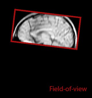
of limiting MWI to the brain, even without the cerebellum, we can now spend about the same amount of time and scan the whole brain and the cervical spinal cord, which is a huge boost for us.” Dr. Rauscher says, “For MWI we perform 3D T2 with 32 or more echoes. This used to take a long time, but with Compressed SENSE we can decrease this to ten minutes for the whole head. Because of the large field of view (FOV) on the readout direction, we even get information from the brainstem, which we previously missed when we were using the GRASE approach. Having the whole head scan is nice because it has spatial resolution, orientation and FOV that are comparable to the standard 3D clinical MS scans, including the FLAIR and 3D T2, and a 3D T1 for brain volume.”
T1 - weighted
Myelin water imaging (echo 1)
With SENSE
With Compressed SENSE
Acquired resolution:
1 x 2 x 5 mm3
→
1.5 x 2 x 3 mm3
Number of echoes:
32 or 48
→
56
Echo spacing:
10 ms or 8 ms
→
7 ms
| | With SENSE | | With Compressed SENSE |
| Acquired resolution: | 1 x 2 x 5 mm3 | → | 1.5 x 2 x 3 mm3 |
| Number of echoes: | 32 or 48 | → | 56 |
| Echo spacing: | 10 ms or 8 ms | → | 7 ms |
T1 - Weighted, Myelin Water Fraction Superimposed
Spinal cord coverage
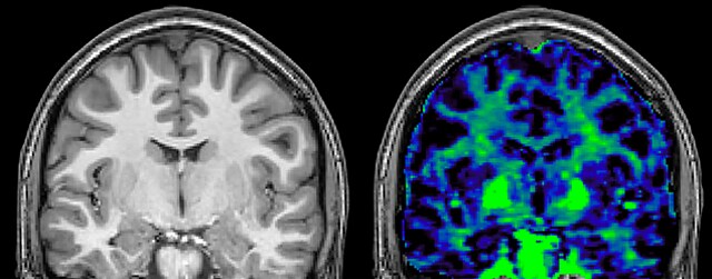
Smaller, more isotropic voxels
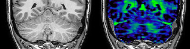
Excellent detail in quantitative maps
Images courtesy of Adam Dvorak, Department of Physics and Astronomy, University of British Columbia

Researchers enjoy flexibility in adjusting sequences, even without pulse programming
The flexibility of the Elition scanner stands out in particular for Dr. MacKay. “Researchers want broad possibilities for manipulating pulse sequences, but getting into pulse programming takes a lot of time and training. Fortunately, the Elition console allows us to do an amazing number of things without going into pulse programming. Technologists or scientists who know the scanner well, can perform scans very quickly, faster than on either of the other scanners we have.” Technologist Laura Barlow agrees: “This system is far more intuitive than our Achieva in terms of navigating operator software; it’s much faster.”
Also Dr. Rauscher considers it a huge advantage that he can simply try things on the Elition that would require pulse programming on different MRI systems, such as their old Achieva system. “For example, adding pre-pulses or turning a conventional 3D T2 scan into an accelerated GRASE scan with multiple echoes have been very easy thanks to the enormous flexibility with the Elition’s graphic user interface,” he says. “In the past, MWI scans were often limited due to time constraints,” says Dr. Kolind, “because it was a difficult acquisition and complicated analysis. But with the Elition, we can perform MWI without having to do any programming modifications.”
"We can now achieve more of our scanning goals in the same time”
Gradient enhancements have a substantial impact on myelin imaging
In practice, Ingenia Elition allows the team to do things now that were not possible before: “There are some scans that we didn’t do before, because the scans were taking too long. With Elition we can combine scans that would have been prohibitively long on the old scanner.
For Dr. Kolind, the Elition excels in advanced neuroimaging for two main reasons. “It's image quality and access to so many different imaging parameters. We’re involved in several multi-center studies, and we can always easily identify the images that came from our Elition scanner, because they are just so beautiful – even though it seems like we’ve set our parameters similarly to other study participants. And as a physicist, being able to do many things, for instance to push resolution and save time, is really helpful.”
According to Dr. MacKay, MWI images benefit from Elition’s high quality gradients. “We need good gradients because we want to be able to do multi-echo sequences that have short TE times.”
Dr. Rauscher says, “With better gradients we can use a shorter echo spacing on the spin echo, so we get better sampling of the rapidly decaying myelin signal, which typically has T2 of around 10-20 milliseconds at 3 Tesla. If we can reduce echo spacing from about 8 to 5-6 milliseconds, we get a much better sampling of the short decay component and increase our SNR, which is a big advantage. The same is true for multi-echo gradient echo which we use for susceptibility mapping and for mapping venous vessels in MS.”
For instance, we can get the combined information from looking at advanced diffusion, multi-shell diffusion combined with myelin water, and then also add quantitative susceptibility mapping and get a better idea of tissue micro structure effects from all three combined. On the old scanner we only had time to run maybe one or two of these sequences in addition to the conventional scans,” says Dr. Rauscher.
Accelerating scans helps researchers achieve their imaging goals
He says the accelerated scanning is achieved via the use of Compressed SENSE and MultiBand SENSE. “We can use Compressed SENSE acceleration factors of about 10 on a 3D FLAIR for instance, which is quite remarkable compared with what we saw with the Achieva. With 3D FLAIR, we can push the spatial resolution to 0.3 cubic mm and it works. Previously, our 3D FLAIR scans lasted about 8 minutes, but now with Elition they are five minutes. The SNR is also visibly better. Our SWI and QSM scans look fantastic. Also, since a lot of neuroimaging is EPI based, using the MultiBand SENSE technique can increase temporal resolution and make it possible to run complicated DTI scans relatively quickly.”
The Elition scanner at UBC has helped researchers decrease scan times and has paved the way for extended scanning possibilities. “We can now achieve more of our scanning goals in the same time,” says Dr. Rauscher. “Patients can typically stay in the scanner for about 45 minutes. So, with the faster scan times, we can now add more scans into one session.
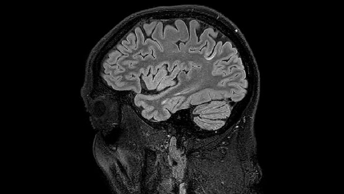
Sagittal 3D FLAIR with 0.3 mm3 voxel volume acquired in 5:12 min. using Compressed SENSE showing a (juxta)cortical MS lesion.
QSM based on a Compressed SENSE multi-echo SWI.
“The progress bar tells patients how much time is left, the little clock shows when to do a breath hold. These may seem little things, but they make a big difference”
Surprised by big impact on patient and volunteer comfort
The great impact of Elition’s patient comfort features was a surprise for Dr. Kolind. “In our research projects we are working with many patients and volunteers. Their universal feedback has really been that it's just such a better experience than the previous scanner.” Dr. Kolind thinks that the comfortable bed and the Ambient system all contribute to patient comfort. “Having control over the lighting and environment with the Ambient Experience seems to really give the patients a feeling that they have more power during the scanning process.”
According to technologist Laura Barlow, many people are quite relaxed when they get off the table. Some explicitly mention that the comfortable mattress and added features, such as ability to see the scan’s progress bar, also keep patients feeling satisfied with their experiences.
“One of our patients admitted to being very uncomfortable in previous MRI scans and had actually tried to avoid having clinical MRIs for four years. She had decreased mobility, so we brought her into the room using the FlexTrak trolley, and she remarked on how comfortable the mattress was. After her scan, she said it was the best MRI she’d ever had, and she would definitely come back and volunteer again.”


Smart features help contribute to a quick workflow for patient preparation and scanning
According to Laura Barlow, the addition of the Elition scanner at UBC has resulted in impressive changes in daily workflow. “It’s really easy to operate the scanner,” she says. “Being able to drag-and-drop, having the middle mouse to scroll through the images, it’s just so much faster than our previous system.”
The staff also notices enhancements in patient preparation prior to scanning. “Bringing patients into the room and positioning them on the table is certainly fast now, since we can move the table without having to landmark with certain coils, and having the FlexTrak makes it easier to help patients on and off the table,” Ms. Barlow says.
“The VitalEye wireless gating system makes things much easier, too. We tested it with volunteers who were instructed to move around during scanning, and it still performed very well, which was a nice surprise. Being able to set the scan to start as soon as we shut the door also improves the flow. The AutoVoice and progress bar cuts down on the amount of time we need to communicate with the patient, which also helps accelerate the scan time.”
“The VitalEye wireless gating system makes things much easier”
An excellent scanner for researchers and their study subjects
“Our decision to purchase Ingenia Elition was based on the image quality and ease of use,” says Dr. MacKay. “We really feel that the images we get from the Elition 3.0T scanner are high quality and more artifact-free than what we see from other systems. Also, the console of the Elition allows our techs and researchers to do an amazing amount of things without having to get into pulse programming. Just sitting at the console, you can do a remarkable number of things with this scanner.”
“What we didn’t foresee beforehand was how much patients would love the scanner,” says Dr. MacKay. “They find it so much nicer to be inside the wide bore Elition with its Ambient and In-bore experience.
They can see the progress bar that tells them how far they are in the protocol and how much time is left, or the little clock when they have to do a breath hold. These may seem little things, but they make a big difference, particularly because patients participating in trials or research studies often have to stay quite long in the scanner.”
“Bringing patients into the room and positioning them on the table is certainly fast now, since we can move the table without having to landmark with certain coils”
Hear the team of the University of British Columbia in Vancouver discuss the benefits that the Philips Ingenia Elition systems brings for their MR research.
Hear from Laura Barlow, lead technologist at the University of Columbia, how she experiences the Philips SmartWorkflow on their Ingenia Elition system.
Summary of experiences with Ingenia Elition at UBC:
References
Results from case studies are not predictive of results in other cases. Results in other cases may vary.
Subscribe to FieldStrength
Our periodic FieldStrength MRI newsletter provides you articles on latest trends and insights, MRI best practices, clinical cases, application tips and more. Subscribe now to receive our free FieldStrength MRI newsletter via e-mail.
Stay in touch with Philips MRI