High quality and fast scanning with Compressed SENSE at KCH
FieldStrength MRI magazine
User experiences - November 2018
Share this article:
At Kurashiki Central Hospital, use of Compressed SENSE resulted in faster and higher quality MRI scans1, which is welcomed by patients and staff
The MRI staff at Kurashiki Central Hospital incorporated Compressed SENSE into most of the brain, spine, abdominal, and cardiac examination protocols on their Ingenia 1.5T. Compressed SENSE accelerates sequences1 without sacrificing image quality. The team realized significant decreases in scanning times, as well as shortened breath hold times that are easier for patients to achieve. In other cases, the acceleration is used for higher spatial resolution or adding an extra sequence. The staff is experiencing increased flexibility thanks to the shorter scanning times, and decreases in workload pressure. After seeing its capabilities on Ingenia 1.5T, KCH decided to acquire Compressed SENSE for two additional MRI scanners.
“After seeing the improvements that Compressed SENSE brought for Ingenia 1.5T, we decided to also acquire Compressed SENSE for two additional MRI scanners”
Takashi Koyama, MD, PhD
Diagnostic radiologist and Director of the Department of Radiology Center and Diagnostic Radiology at Kurashiki Central Hospital. His focus is on diagnostic imaging in general radiology, especially genitourinary MRI.
Masayuki Kumashiro, PhD
Radiological technologist and Director of Radiological Technology Kurashiki Central Hospital. He is Vice President of Japan Association of Radiological Technologists. He was member of the Ministry of Health Committee of State Examination for Radiological Technologists in 2008-2016. His specialties include cardiovascular and neurovascular MRI and IVR.
Koshi Miyake, MD
Cardiologist and Chief Physician in Department of Cardiology at Kurashiki Central Hospital. He is a Board certified member of the Japanese Circulation Society.
Sachi Fukushima, RT
Radiological technologist and member of the MR technical staff at Kurashiki Central Hospital. She is a certified technologist, and an authorized magnetic resonance technological specialist.
Improvements in scanning without sacrificing image quality
At Kurashiki Central Hospital (KCH) in Japan, the MRI staff is always looking for ways to improve image quality and to scan faster. As one of the goals of faster scanning, they mention improving the MRI examination for patients, for instance by reducing breath hold duration and overall scanning time. This can make it easier for patients to comply, which in turn benefits the imaging results. An additional aim is to gain time for the MRI technologists, to relieve the pressure of their high workload and to give them more time to attend to their patients.
When the staff at Kurashiki heard about Compressed SENSE, they were eager to learn about its capabilities, and what these could mean for their staff workload and patient comfort. KCH is a general hospital that scans approximately 110 patients a day using seven MRI systems. Compressed SENSE was first installed on their Ingenia 1.5T scanner. The staff quickly appreciated substantial decreases in scan times and decreases in breath hold requirements, without having to compromise on image quality.
“With Compressed SENSE we can obtain excellent quality images even when using higher C-SENSE factors for decreasing scan times”
Convincing results driving quick adoption
According to KCH diagnostic radiologist Dr. Takashi Koyama, Compressed SENSE is extensively used in brain and spine examinations. Both of which require high quality images, and it is possible to obtain excellent quality images even when using higher Compressed SENSE factors (C-SENSE factors) for decreasing scan times. Brain protocols were the first to be converted to Compressed SENSE, according to MRI technologist Sachi Fukushima. “We started with comparing the image quality of Compressed SENSE with our original SENSE images in five cases of brain imaging. For 2D images for instance, we looked for possible changes in contrast and structural details when changing from a SENSE factor of 2.0 to a C-SENSE factor of 2.4. We also examined image quality obtained with different denoising parameters.” This convinced the Kurashiki team that image quality would not suffer by switching from SENSE to Compressed SENSE.
The Compressed SENSE technology allows users to accelerate their 2D and 3D sequences by up to 50%.1 The staff at KCH has noticed reduced noise in many Compressed SENSE images in different anatomies, allowing them to increase the spatial resolution. “We are now using Compressed SENSE for almost all of our sequences in brain, spine and abdominal examinations,” says technologist Masayuki Kumashiro, PhD.
“We first compared image quality before switching from SENSE to Compressed SENSE”
Fast brain scanning allows to include more high resolution 3D sequences
“For several brain examinations we use Compressed SENSE for faster1 scanning, using C-SENSE factors from 2.0 to 3.5 for 2D and 3D protocols (except EPI diffusion). High resolution sequences such as TOF and 3D usually take the longest, but with Compressed SENSE we obtain really good quality, even when using a higher C-SENSE factor for decreasing scan time as compared to images with SENSE.” “In the 3D TOF MR angiography sequence, we decided to use a C-SENSE factor of 3.5, after carefully examining the quality of the inflow signal and the detailed structure of the cerebral blood vessels using this higher factor. Compressed SENSE also allows us to use more 3D sequences instead of 2D sequences, providing higher resolution and allowing us to perform reformatting (MPR) in different orientations,” says Fukushima.
According to Fukushima, the high spatial resolution desired in brain and spine, used to typically require quite long scan times, but now the speed provided by Compressed SENSE may be traded for increased spatial resolution when necessary.
“Because the faster scanning with Compressed SENSE saves us time, we can add a sequence to obtain high quality spine images for confident diagnoses”
Boosting diagnostic confidence with multiple contrasts, multiple orientations in spine MRI
“Previously with SENSE, our 2D mDIXON TSE scans required relatively long scan times. But now, with Compressed SENSE, we have reduced these scan times while maintaining a high SNR, because the Compressed SENSE technology helps reduce noise,” says Dr. Koyama. “Because the faster scanning with Compressed SENSE saves us time, we can sometimes add a sequence to obtain high quality spine images in the same time slot for confident diagnoses. And in cervical spine exams, a 2D sequence is sometimes replaced by a 3D protocol, which provides us more information as it can be reformatted in different orientations. Compressed SENSE allows us to easily add this 3D sequence in the timeslot,” says Fukushima “Incorporating Compressed SENSE in common spine sequences, such as mDIXON, 3D SpineVIEW and eTHRIVE, can substantially reduce the scanning time of these sequences, while maintaining adequate spatial resolution, resulting in high quality, multiple contrasts, multiple orientations,” says Dr. Koyama. “In addition, fast sequences generally make it easier for patients to stay motionless throughout the scans, so it also helps us in that way.”
The KCH team has already changed most of their Ingenia 1.5T spine ExamCards by incorporating Compressed SENSE into their 2D TSE, mDIXON TSE, FFE, and 3D sequences.
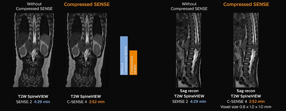
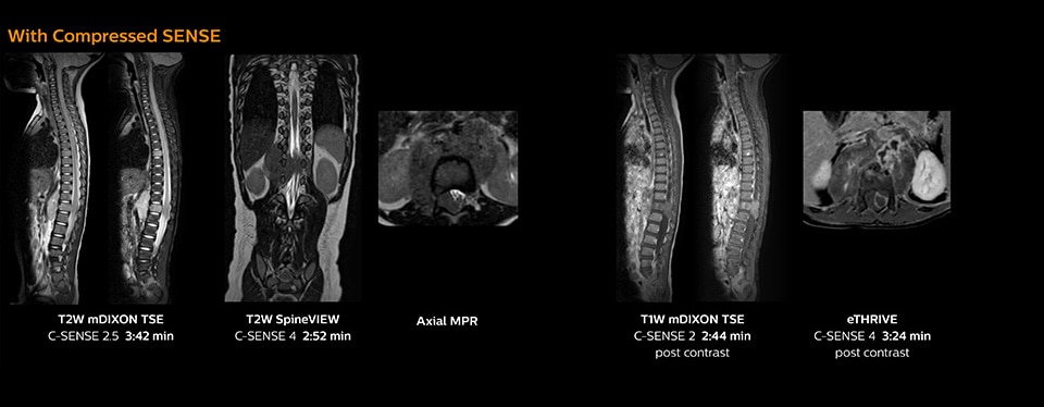
Pediatric spine with neuroblastoma
This 6-year-old patient with neuroblastoma underwent MRI on the Ingenia 1.5T. Compressed SENSE was used to reduce scan time while maintaining the high resolution for 2D mDIXON, 3D SpineVIEW and e-THRIVE in this case. The highly detailed images allowed the radiologist to make a quick and confident assessment of the position of the nerve and the tumor. Especially important for a pediatric patient, is that a shorter scan time also allows us to keep the sedation time as short as possible.
As this was one of the first patients scanned with Compressed SENSE, 3D SpineVIEW was acquired with and without Compressed SENSE to allow comparison. Although the Compressed SENSE sequence was significantly faster, the acquired and reconstructed 3D SpineVIEW images show virtually the same image quality.
View ExamCard on NetForum: Ingenia 1.5T Pediatric spine with Compressed SENSE – Kurashiki Central Hospital
“Compressed SENSE has allowed us to increase spatial resolution in breast MRI, which benefits our diagnostic confidence”
Identification of small breast lesions requires high resolution
“In breast scanning, high resolution is important to help me identify very small mammary lesions, so, we need high spatial resolution in 2D T1- and T2-weighted images, as well as a short scan time. Compressed SENSE has allowed us to increase spatial resolution, which benefits our diagnostic confidence.”
Dr. Koyama says that he used to believe that high resolution MRI at 1.5T required long scan times, and SNR was low. “With Compressed SENSE, however, it is possible to acquire high quality images, even with higher Compressed SENSE factors, so in a quite short time.”
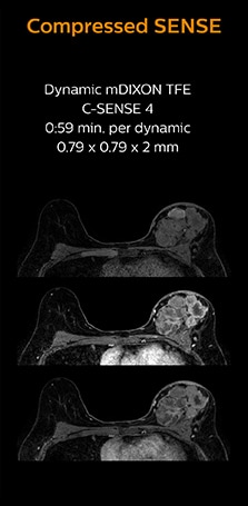
Dr. Koyama says that a C-SENSE factor of 4.0 was chosen to increase their spatial resolution in 3D dynamic breast scanning. “In addition to a high temporal resolution, we also require high spatial resolution, which helps us to see details of the internal structure of the lesion and to see lesions separately from normal anatomic structures. We can also see if a lesion extends into adjacent organs and anatomic structures.”
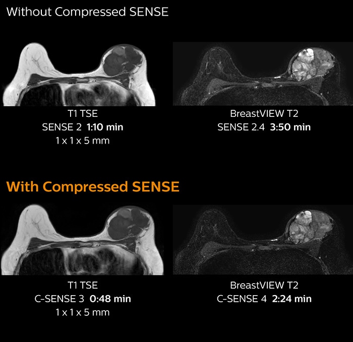
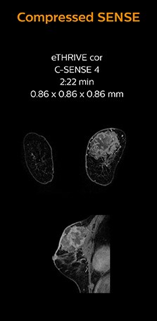
3D MRI with Compressed SENSE of patient with breast cancer
This patient underwent MRI on Ingenia 1.5T with Compressed SENSE. Compressed SENSE was used to reduce the scan time in order to decrease the time that the patient may experience discomfort and pain, both of which may lead to patient motion. The 3D BreastVIEW and 3D high resolution mDIXON images – both mDIXON contrasts are acquired in the same exam – show high quality images even with the shorter scan time. The spatial resolution of the dynamic scan with Compressed SENSE is higher than in the previous protocol (not shown) which allows for better visualization of the lesion with respect to the muscles of the thoracic wall and better delineation of small structures.
For breast imaging, a fast, high resolution scan can be important for a female patient having to lie in an uncomfortable, face-down position in the scanner. Compressed SENSE also helps us to obtain higher1 quality images using 3D BreastVIEW and 3D high resolution mDIXON sequences in the same examination time as in our previous exam protocol.
View ExamCard on NetForum: Ingenia 1.5T Breast with Compressed SENSE – Kurashiki Central Hospital
“Since short breath hold times make it easier for our patients to comply, the failure of breath holding largely disappeared”
Shortening scan times to reduce breath hold failures in abdominal MRI
KCH technologist Fukushima recognizes the impact of Compressed SENSE on breath hold times. “Before we started using Compressed SENSE, some high resolution 3D examinations used to require long breath holds, because when scan times were shortened, images would often become noisy in the center of the body. But now, Compressed SENSE allows us to further decrease scan times without that increased noise.” Dr. Koyama particularly focused on reducing breath hold times in abdominal MRI, as these patients often need to perform many breath holds in one examination. “In liver scans we were able to shorten our scanning time – and thus reduce breath hold time – from 22 to 13 seconds by using a higher C-SENSE factor. Since shorter breath hold times make it easier for patients to comply, we saw breath holding failures largely disappeared, allowing the exam to be finished smoothly.”
While breath holds are vital for image quality in many abdominal MRI scans, commonly used breath hold times of around 25 seconds or more can be challenging, for instance when a patient is sick, in pain or stressed. Failed breath holds can decrease image quality and add to scan time.
“In our abdominal EOB examination, we previously used a SENSE factor of 2 in T2w TSE with a scan time of 24 seconds in one breath hold. But now, C-SENSE factor 2 helped us reduce this to only 16 seconds. And the 3D eTHRIVE changed from SENSE factor 3 with 20 seconds breath hold to C-SENSE factor 5 with only 16 seconds breath hold time.
Faster1 cardiac imaging with fewer or shorter breath holds
“Compressed SENSE has now been implemented in all cardiac exams. Thanks to the acceleration, fewer breath holds are now needed, or breath hold times are shortened. This reduces the burden of the exam for cardiac patients, without affecting the quality of information required for cardiac function analyses,” he says. “Because it’s easier for patients to comply with the breath hold times” “In our previous cardiac cine sequence, we were acquiring two slices during one breath hold. With Compressed SENSE, we increased this to four slices per breath hold. It is also possible to shorten scanning time using a C-SENSE factor 6 without sacrificing image quality.”
Dr. Koshi Miyake, cardiologist, explains that scanning of patients with cardiac arrhythmia can be challenging, as scanning times can become very long due to the varying heart rate. So his most important motive to implement Compressed SENSE in cardiac MRI exams was to reduce the burden of breath holds for the patient, while maintaining high image quality. He hopes this can also help to reduce motion caused by the difficulty for patients to hold their breath.
“In cardiac MR we can now increase from two to four slices per breath hold”
Making Cardiac MR faster and easier for patients
“In cardiac MR fewer breath holds are now needed, or breath hold times are shortened”
“Typically, in scans with high contrast, such as 2D balanced TFE cine, a quite high C-SENSE factor may be used. When we tried a higher C-SENSE factor, we saw still no significant influence on cardiac ejection fraction, but 2D image quality started to decline. For coronary imaging, we use a C-SENSE factor of 3 in 3D balanced TFE, or even up to 4 when contrast is high.”
KCH to soon extend Compressed SENSE to more scanners
The KCH staff would definitely recommend Compressed SENSE to peers at other institutions. They advise new users to start using it in brain and spine imaging first, as these are the easiest protocols to switch. According to Dr. Kumashiro, “The most important reason for us to acquire Compressed SENSE has been to achieve higher image quality and make examinations easier for patients by reducing breath hold scan time and examination time. The shorter scan times achieved with Compressed SENSE relieve the stress of a tight work schedule for the MR staff. Technologists can spend the gained time on increasing the image quality, or to take more time for patient preparation and dealing with safety aspects.” “After seeing the improvements that Compressed SENSE brought to the scanning on Ingenia 1.5T, we decided to also acquire Compressed SENSE for two additional MRI scanners,” he concludes.
“After seeing the improvements that Compressed SENSE brought to the scanning on Ingenia 1.5T, we decided to also acquire Compressed SENSE for two additional MRI scanners”
“Because Compressed SENSE results in shorter scan times, this relieves the stress of a tight work schedule for our MR staff”
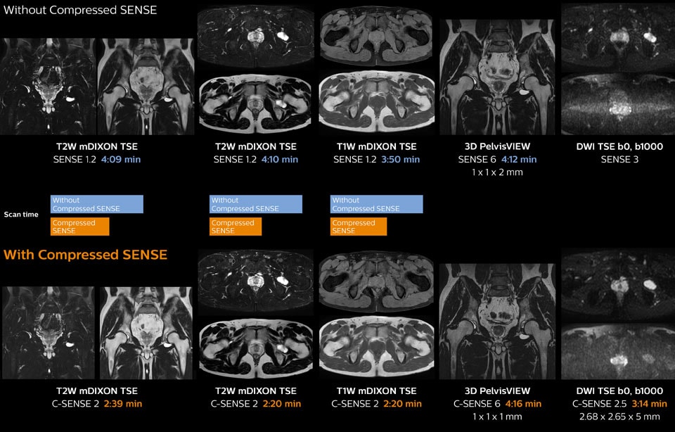
Hip with synovial cyst w/wo Compressed SENSE
Ingenia 1.5T MRI scans with and without Compressed SENSE are compared for this case of a hip with a synovial cyst. In the mDIXON sequence, the Compressed SENSE factor used is higher than the SENSE factor on the previous scan, so scan times are reduced without sacrificing image quality. The high resolution images are useful for diagnosing of a detailed dissection. The diffusion TSE with Compressed SENSE shows reduced noise compared to the Diffusion TSE with SENSE and the artifact has disappeared.
So in this case, Compressed SENSE helped in reducing scan times of the examination. The Compressed SENSE images allowed the radiologist to confidently diagnose the lesion and see the anatomic relationships of the abnormal signal to the surrounding structures.
View ExamCard on NetForum: Ingenia 1.5T Hip with Compressed SENSE – Kurashiki Central Hospital
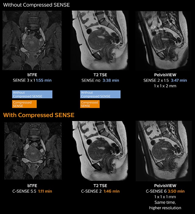
Pelvis with myoma – comparison w/wo Compressed SENSE
In this MRI exam of pelvis in a patient with myoma, Compressed SENSE is used to accelerate individual sequences and thus the entire examination on Ingenia 1.5T. Compressed SENSE allowed for a decrease in scan time for the T2 TSE from 3:38 to 1:46 minutes. The Compressed SENSE images in this case show fewer motion artifacts than the images from the previous protocol with SENSE. In 3D PelvisVIEW, the Compressed SENSE images have a higher and isotropic spatial resolution with a scan time similar to the SENSE sequence. The improved spatial resolution and better contrast in the myometrium of the uterus allowed radiologist Dr. Koyama to confidently diagnose the cancerous lesion in the uterus. The use of Compressed SENSE accelerates scanning times and increases spatial resolution in 3D PelvisVIEW.
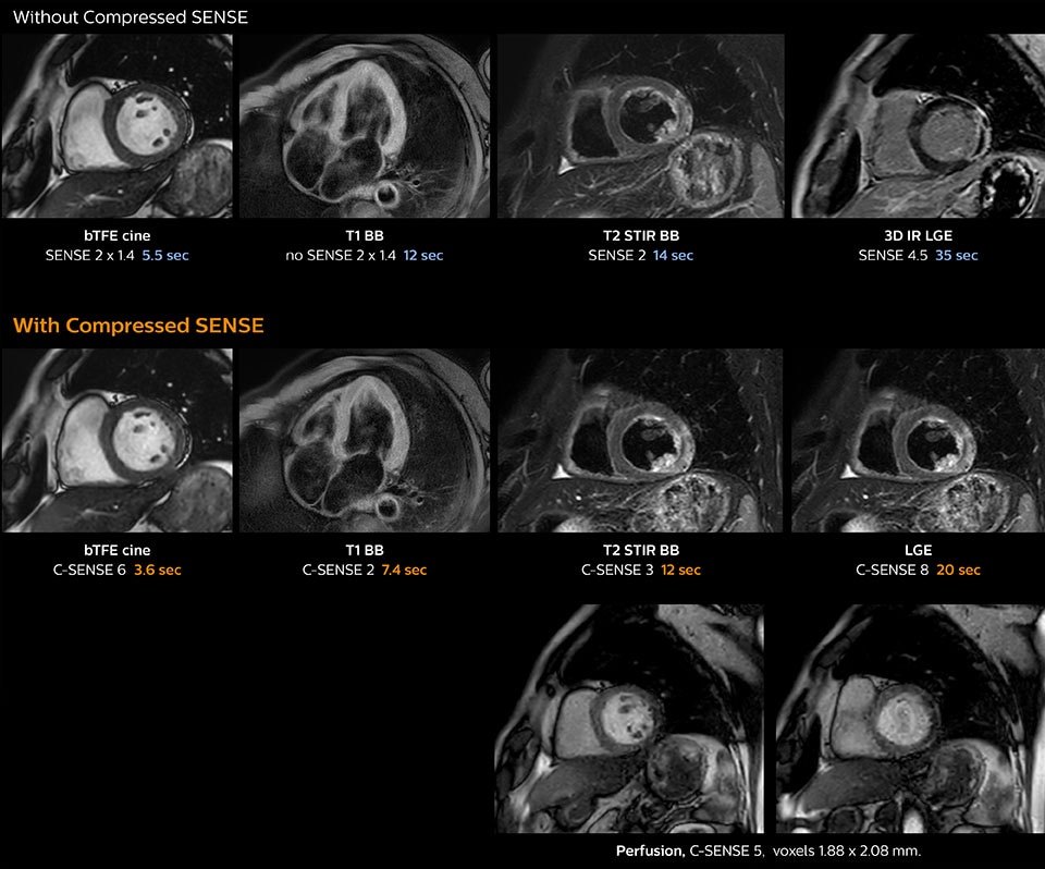
Cardiac MRI of acute myocardial infarction (AMI) w/wo Compressed SENSE
These images of a patient with acute myocardial infarction images were acquired on Ingenia 1.5T with and without Compressed SENSE.
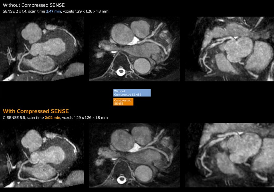
3D whole heart MRI and coronary arteries w/wo Compressed SENSE
These images with and without Compressed SENSE were acquired on Ingenia 1.5T.
View ExamCard: Ingenia 1.5T Cardiac imaging with Compressed SENSE - Kurashiki
1Compared to examinations without Compressed SENSE.
Results from case studies are not predictive of results in other cases. Results in other cases may vary.