
Scan faster* and have more time for advanced MRI techniques
FieldStrength MRI magazine
User experiences – April 2020
Fondation Rothschild creates more time for advanced neuro exams by faster* scanning, thus boosting diagnostic confidence
When faced with the task of selecting two MRI scanners for a busy neurology clinic, Dr. Julien Savatovsky and his team looked for a system that could help them meet the increasing demand for MRI exams, and deal with declining reimbursements in France. After a balanced assessment of several systems, the quality of the images and the fast acquisition times of Ingenia Elition 3.0T led to the decision to purchase two Elition scanners. These have allowed the team to shorten scan times, and that enables Dr. Savatovsky to include more advanced sequences to boost diagnostic confidence.
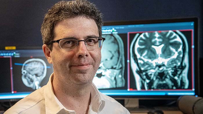
Julien Savatovsky, MD
Diagnostic neuroradiologist at Fondation Rothschild, Paris, since 2006. His clinical interests include advanced diagnostic imaging in neuro, head and neck, and ophthalmology.
“Many examinations are shortened, so for more complex cases we can add advanced sequences or switch to higher resolution for improving diagnostic confidence”
The need for flexibility while maintaining a steady workflow
Fondation Rothschild, based in Paris, France, is a tertiary care hospital that performs specialized workups for patients presenting with neurologic, ophthalmologic, and head-and-neck diseases. They also provide first-line treatment for patients, as well as emergency care for acute stroke, ophthalmologic, and neurologic cases. The radiology department has been using four MRI scanners, including two Ingenia Elition 3.0T machines, since November 2018. According to Dr. Julien Savatovsky, a diagnostic neuroradiologist at Fondation Rothschild, the increasing demands for MRI exams, decreasing reimbursements in France, and the need to perform advanced exams for complex patients requires fast, high quality scanning and efficient workflow. These challenges drove the decision to buy two Ingenia Elition 3.0T scanners. From the beginning, the intention was to acquire two of the same machines. “We like the two MRIs to be interchangeable, so that we can easily switch a patient to the other device when we have to accommodate emergencies during the day”, says Dr. Savatovsky. “Another reason is that this allows our technologists to use both devices in exactly the same way and they only need to learn one user interface.”
Image quality and speed driving the Elition purchase
The choice for two Elition scanners was not made lightly. Dr. Savatovsky and his team compared devices from different vendors before making the decision. “Part of this process was our assessment of the image quality of different devices. We put together a list of sequences with detailed requirements, including limited acquisition times, to allow a fair comparison. We even put the same volunteer in each scanner. Our assessment was that the image quality was better with the Elition scanner. Compressed SENSE or Multiband SENSE was used for almost every sequence, and I think this helped a lot to maintain a great image quality in the shorter acquisition time.”
“The main breakthrough for us was that Compressed SENSE and Multiband SENSE have allowed us to accelerate our examinations.”
Using speed for shorter exams or more information in the same acquisition time
According to Dr. Savatovsky, Ingenia Elition has an impact in virtually all examinations. “We can either make the scanning faster compared to our older Ingenia 3.0T, or we save enough time so that we can add sequences we wouldn’t perform otherwise, or increase resolution. So, I think it has benefits for most of our patients.” “Some routine exams that we use every day have been shortened since we started using Elition. For example, we now use mostly a comprehensive stroke protocol (high b-value diffusion, fast 3D FLAIR, TOF, supra-aortic vessels angiography, SWIp, T1 post gad) that lasts 10 to 11 minutes, but our fast stroke protocol takes only 7 minutes. Our routine IAC needs about 10 minutes scan time and our comprehensive brain MS examination requires no longer than 13 minutes of scan time. Our ability to reduce acquisition times of most sequences helps to shorten total examination times, which in turn helps us to increase the number of patients we scan per day. “The main breakthrough for us was that Compressed SENSE and Multiband SENSE have allowed us to accelerate our examinations. Alternatively, we can invest the time gained in obtaining higher spatial resolution to see more details, or we can add additional sequences,” says Dr. Savatovsky. “That’s a big improvement from what we did before.”
“We can invest the time gained in obtaining higher spatial resolution to see more details, or we can add additional sequences. That’s a big improvement from what we did before."
Improving scan time and/or spatial resolution
A two year follow-up scan of a CPA meningioma using both Ingenia 3.0T without CS and Ingenia Elition 3.0T with Compressed SENSE allows for a nice comparison to demonstrate the protocol improvements achieved on the Elition: 3D FLAIR has a shortened scan time, improved SNR and still the same spatial resolution. BrainView (3D T1 TSE) has improved spatial resolution and SNR with shortened scan time. For 3D T2 Drive the spatial resolution has been improved. 3D THRIVE used to have an interpolated 0.8 mm slice thickness, but true thickness at 1.6 mm, so that axial slices displayed a decent quality, but reformats were suboptimal. Compressed SENSE is used on Elition to improve spatial resolution and reduce the non-interpolated slice thickness to allow smoothly reformatted images. Total scan time (adding SmartBrain and an additional b2000 diffusion) was 13:19 on Ingenia, and is now reduced to 10:42 on Ingenia Elition.
Ingenia 3.0T (without Compressed SENSE)
3D FLAIR 1.0 x 1.0 x 1.0 mm* 4:24 min.
3D TSE T1w 1.0 x 1.0 x 1.2 mm* 2:40 min.
3D T2w Drive 0.8 x 0.8 x 1.0 mm* 3:05 min.
3D T1w THRIVE 0.8 x 0.8 x 1.6 mm* 1:30 min.
Ingenia Elition 3.0T with Compressed SENSE
3D FLAIR 1.0 x 1.0 x 1.0 mm* 2:50 min.
3D TSE T1w 1.0 x 1.0 x 1.0 mm* 2:10 min.
3D T2w Drive 0.7 x 0.7 x 0.7 mm* 2:52 min.
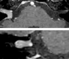
3D T1w THRIVE 0.7 x 0.7 x 0.8 mm* 1:30 min.
*true voxel size, without interpolation
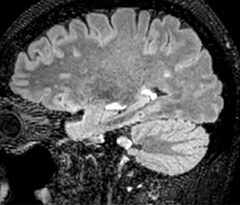
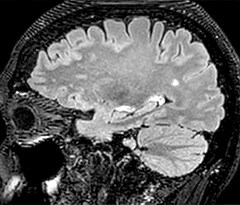
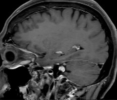
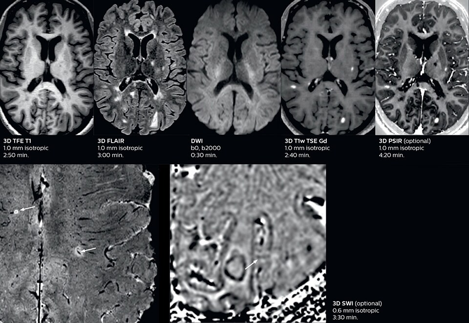
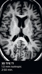
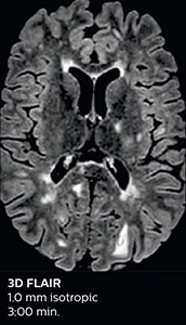
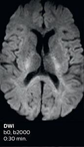
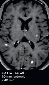
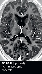
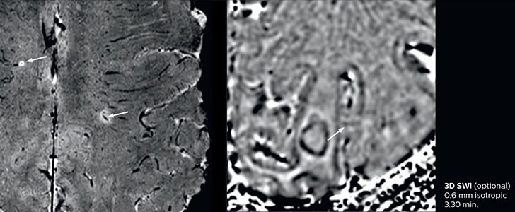
The abbreviated MS protocol for brain is only around 9 minutes, so in case of suspected multiple sclerosis, one or two more advanced sequences may be added, such as PSIR (phase sensitive inversion recovery) or susceptibility-weighted sequences to help us make more confident diagnoses in these inflammatory cases.
In this example, the optional 3D multishot susceptibility weighted sequence with 0.6 mm isotropic voxels is 2 lesions with a central vein sign (arrows) and one lesion with a phase-rim sign (arrowhead). The total scan time, including SmartBrain and axial PD/T2 3mm, is 11:10 min. and is 18:30 min. with the optional 3D PSIR and 3D SWI multishot included.
“In patients that need a classification for brain mass, we can provide a more detailed and confident diagnosis than before”
Enriching examinations to boost diagnostic confidence
“We used to have long examination times for certain types of patients, a few lasting more than 40 minutes,” says Dr. Savatovsky. “What is remarkable, is that now all these examinations are below 30 minutes, which opens up opportunity to add more sequences when needed. It’s really hard to keep a patient for more than 40 minutes in the scanner, but because we have now cut scan times by at least 10 minutes, we can add more sequences without making the exam too long. And this is where the new system helps us make a difference. Examples include our examinations for informing brain tumor classification or giant cell arteritis workup, or for intracranial wall imaging – so in patients where we need several advanced sequences or high resolution sequences.” “We added three additional sequences in our brain neoplasm classification exam: a 3D SWI sequence, APT and ASL on top of 3D morphologic sequences, an isotropic DSC (dynamic susceptibility contrast) and multivoxel spectroscopy. I think that in patients that need a classification for brain mass, for example, we can provide a more detailed and confident diagnosis than before, allowing the clinicians to decide for either a medical workup if no tumor is suspected, or for neurosurgery as soon as possible if a neoplasm is suspected.” “In multiple sclerosis patients, we increasingly include a multishot susceptibility sequence [3] in our routine cases, thanks to the shorter scan times. Our abbreviated MS protocol for brain is around 8 to 9 minutes, so we can ask for one or two additional sequences to visualize the central veins, or to get an additional contrast to better depict posterior fossa lesions. In cases of white matter lesions of unknown significance on FLAIR images, for example when we see high signal hyperintensities in the brain, we can add on more advanced sequences such as PSIR (phase sensitive inversion recovery) or susceptibility-weighted sequences to help us in distinguishing between MS and nonspecific or vascular abnormalities in these inflammatory cases.”
Performing advanced techniques has a direct impact on hospitalizations
So, what is the actual impact of having more information and more diagnostic confidence? According to Dr. Savatovsky, “One of the indications I’ve seen where using Elition is most impactful is in patients with suspected giant cell arteritis. As an ophthalmologic hospital, we see many patients with suspected giant cell arteritis. Usually we were performing MRI to help us rule out an ischemic stroke, and to verify that the supra aortic vessels are undamaged. With Elition, we still do this, but now we can add on more detailed high-resolution black-blood sequences on superficial arteries. This provides us with high confidence levels for diagnosis of giant cell arteritis (GCA) and as a result, some patients are not sent for a biopsy anymore. A patient who has a normal MRI will not require a biopsy and can be discharged from the hospital in the same day. Before, such a patient would have to stay for about a week, just to find that their biopsy results were negative. We have at least three or four patients a week with suspected giant cell arteritis. For a great deal of these patients we can have a direct impact on their hospital stay.”
Hear Dr. Savatovsky discuss about the benefits that the two Ingenia Elition systems are bringing their clinical practice.
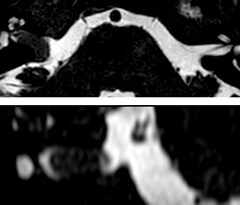
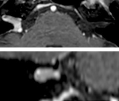
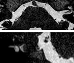
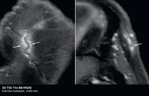
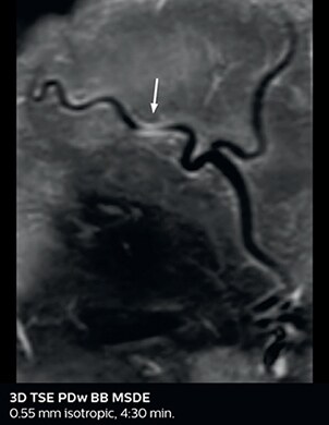
Giant cell arteritis
The 3D TSE T1w black blood MSDE sequence with fat suppression has an isotropic 0.8 mm voxel size and sagittal oblique and axial reformats are made. The images show superficial temporal artery thickening and peri-arterial fat infiltration. The 3D TSE PDw black blood MSDE with fat suppression has 0.55 mm isotropic voxels. The images shows focal involvement of the frontal branch of the superficial temporal artery.
A good workflow for excellent patient management
Dr. Savatovsky recognizes that throughput in an MRI center not only depends on scanning time, but also on the time needed by the technologist to position the patient and operate the MRI scanner. “Scan time in very short exams will last from 5 to 9 minutes. Our goal is to keep our average scan time under 15 minutes.” “The time needed for positioning the patient and the technologist’s workflow depends on the device. We particularly benefit from the lightweight FlexTrak dockable patient transport system for patient preparation outside the MRI room, which we purchased with the Elition. Currently, patient positioning is quite fast, even for heavy or bedridden patients, because with FlexTrak the patient and coils can be installed in the preparation room and then be brought into the scanner room immediately after the previous examination ended.” Other Elition features that support a fast workflow include the auto-start function that starts the scan as soon as the technologist closes the scanner room door.
“The fast scanning capabilities that came with Elition allow us to do a really quick examination and answer a lot of questions within a short time."
Accommodating multiple emergencies per day
The MRI center at Fondation Rothschild receives several neuro and head/neck emergency cases per day. On weekdays, an average of 7 unscheduled patients will require scanning, with approximately 4 to 5 patients actually requiring an urgent MRI scan, according to Dr. Savatovsky. He notes that the ability to accelerate sequences while maintaining image quality is particularly important in the emergency setting. “The fast scanning capabilities that came with Elition allow us to do a really quick examination and answer a lot of questions within a short time. We use every tool available to accelerate image acquisition while maintaining a reasonable image quality. So, for most of the sequences we use Compressed SENSE, for example, in our 3D FLAIR, in contrast-enhanced and noncontrast MR angiography, and for susceptibility-weighted sequences.” Among the emergencies that are routed to the MRI department at Fondation Rothschild, stroke is seen almost daily. “After arriving, acute stroke patients are immediately brought to the MRI preparation room and positioned on the FlexTrak table. There, the neurologist examines the patient and the biological workup is performed. Once this is finished, we can immediately move the patient with FlexTrak into the MRI and begin the scanning within one or two minutes. So, having the FlexTrak is a big advantage for us.”
“For stroke, it allows us to cut about 5 minutes off of our stroke protocol, or to keep the same acquisition time and get more insights”
Comprehensive stroke MRI within acceptable time
Dr. Savatovsky appreciates the improvements and flexibility that Elition with Compressed SENSE and MultiBand SENSE provides, particularly for stroke patients. “For stroke, it allows us to cut about 5 minutes off of our stroke protocol, or to keep the same acquisition time and get more insights.” The ability to perform more sequences can help in making a swift and confident diagnosis. “For example, our stroke cases usually include the regular sequences that every center does (b1000 diffusion, FLAIR, time-of-flight angiography), but we also image supra aortic vessels, and we can replace a gradient echo sequence with a fast 50-second susceptibility-weighted sequence, and all of this doesn’t add much time. because all the regular sequences are accelerated on Elition.” “The time savings with Compressed SENSE and MultiBand SENSE make it easier to add sequences to give us additional insights. Depending on the context and the first results, we might add a DSC perfusion to assess the ischemic penumbra, an ASL perfusion to help find an alternative cause in case of normal diffusion, or add a high-resolution T1 sequence for a stroke patient, to quickly assess wall imaging in emergency cases. The additional sequences can help improve patient management, because we can already consider some alternative diagnoses if the morphological MRI is normal.”
Improved diffusion imaging in stroke patients
Using MultiBand SENSE allowed the staff to improve their diffusion quality. “Our diffusion sequence was already fast before, about 40 seconds. Now with Elition, it still lasts 40 seconds, but we improved the spatial resolution by 0.2 mm and use high b-values to be more sensitive to visualize changes related to acute stroke,” says Dr. Savatovsky. “We now also developed a high resolution DTI sequence (1.3 x 1.3 x 2 mm) that can be reformatted and takes 2 to 5 minutes depending on the coverage. We use it every time we have a doubt, or when we expect the diffusion to be abnormal but don’t see that on the fast sequence. We occasionally spot small ischemic infarctions that would not have been visible with the regular diffusion sequence.”
“We now also developed a high resolution DTI sequence (1.3 x 1.3 x 2 mm) that can be reformatted and takes 2 to 5 minutes depending on the coverage.
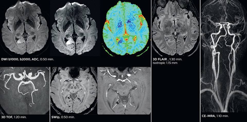
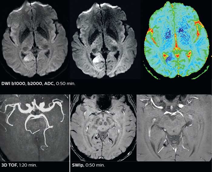
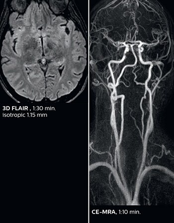
Fast acute stroke protocol
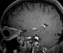
This is an example of acute ischemic stroke with distal occlusion of the right posterior cerebral artery. Note the improved visibility of the ischemic territory on the diffusion weighted image with high b-value. The 3D FLAIR shows a distal PCA occlusion. The fast SWIp depicts the thrombus on the isolated second echo image. The total scan time (including SmartBrain, preparations and a fast 3D T1w TSE Gd) is 8:00 minutes.
Excellent spine and spinal cord imaging
Looking for inflammatory lesions of the spinal cord is usually challenging with MRI, says Dr. Savatovsky. “We solved some of the challenges by implementing sequences such as 3D PSIR, which allows us to see far more lesions than the usual T2 imaging. We are starting to see cases where the MRI images at 1.5T were normal, but then we do see lesions when performing the PSIR at 3.0T.” [1] “Elition also performs very well in imaging of the bony spine, the discs and degenerative disease, especially now that we can include at least one 3D sequence in every scan. For example, we perform a lot of 3D spin-echo (TSE) sequences when imaging degenerative lumbar spines. Thanks to Compressed SENSE and the 3D SpineVIEW protocols, we have a very high signal intensity with no flow voids, so the image quality is very good. The possibility to reformat the images in every plane raises the diagnostic confidence, especially in patients who have to undergo surgery.”
Increased patient throughput with higher resolution
Previously, about 30 patients a day were scanned on their Ingenia, 3.0T, during a 15-hour opening period. With the Elition system, the average number of patients scanned per day increased by approximately 10%. “On a day where we only scan outpatients, we can scan 35 to 40 patients a day. Also, routine MRI cases, such as IAC, headaches workup, memory impairment, acute neurological deficit, and multiple sclerosis, last less than 15 minutes, which is appreciated by patients who prefer short examination times,” says Dr. Savatovsky. “Elition really makes a difference. For example, sequences that lasted 6 minutes a few months ago are now completed in 3.5 minutes.”
Purchasing Elition was a wise decision
In conclusion, Dr. Savatovsky recaps the advances made possible by having the Elition scanners: “Many examinations are shortened, for more complex cases we can add advanced sequences or switch to higher resolution for improving diagnostic confidence. Emergency patients in particular benefit from the speed and efficient workflow associated with the FlexTrak patient transport system.” “The image quality for neurological cases is the most robust we’ve seen. If we compare examination time and image quality of a given sequence, like 3D TSE FLAIR, there is no question that the Elition performs really well. We are now also capable of performing multishot susceptibility and can perform PSIR sequences for spine and brain, which are not available on all systems. We can use Compressed SENSE for every sequence, whereas with other vendors this might be limited to a few sequences, only.” Looking back at the economic factors that contributed to his hospital’s decision to purchase two Elition MRI devices, he notes: “Since reimbursement costs are decreasing, we had to calculate the number of patients needed to make an economically sound investment. Now we know that also from an economic perspective, purchasing the Elition MRI machines was a wise decision.”
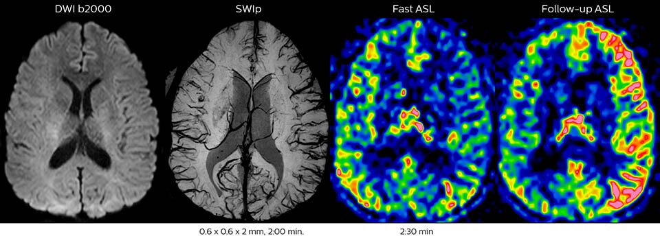
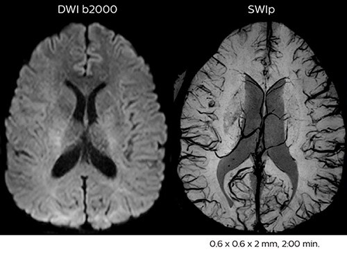
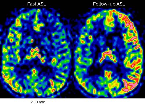
Acute right motor deficit and aphasia
In this patient with acute right motor deficit and aphasia, the b2000 diffusion weighted image is normal. The SWIp image demonstrates more prominent veins in the right hemisphere, which could reflect increased deoxyhemoglobin contents. Fast ASL shows low CBF regions in the left frontal lobe. A follow-up ASL after one hour demonstrates high CBF values in the same area. The final diagnosis was migraine with aura.
Summary of Dr. Savatovsky’s experiences with Ingenia Elition 3.0T:
* Compared to Philips scans without Compressed SENSE ** Compared to the Ingenia 3.0T
References
[1] Mirafzal S, Goujon A, Deschamps R, Zuber K, Sadik JC, Gout O, Lecler A, Savatovsky J.. 3D PSIR MRI at 3 Tesla improves detection of spinal cord lesions in multiple sclerosis. J Neurol. 2020 Feb;267(2):406-414. doi: 10.1007/ s00415-019-09591-8. Epub 2019 Oct 26. [2] Mohammed-Brahim N, Clavel G, Charbonneau F, Duron L, Picard H, Zuber K, Savatovsky J, Lecler A. Three Tesla 3D High-Resolution Vessel Wall MRI of the Orbit may Differentiate Arteritic From Nonarteritic Anterior Ischemic Optic Neuropathy. Invest Radiol. 2019 Jul 16. doi: 10.1097/RLI.0000000000000595. [Epub ahead of print] PMID:31335635 [3] Sati P, et al; NAIMS Cooperative. The central vein sign and its clinical evaluation for the diagnosis of multiple sclerosis: a consensus statement from the North American Imaging in Multiple Sclerosis Cooperative. Nat Rev Neurol. 2016 Dec;12(12):714-722. doi: 10.1038/nrneurol.2016.166. Epub 2016 Nov 11. Review.
Results from case studies are not predictive of results in other cases. Results in other cases may vary.
Subscribe to FieldStrength
Our periodic FieldStrength MRI newsletter provides you articles on latest trends and insights, MRI best practices, clinical cases, application tips and more. Subscribe now to receive our free FieldStrength MRI newsletter via e-mail.
Stay in touch with Philips MRI