HeartNavigator

Insightful planning and guidance for Structural Heart Disease Procedures

Caută produse similare

Increase confidence and ease during transcatheter aortic valve replacement (TAVR) and other challenging SHD procedures. The immersive user experience is highly automated to simplify planning, device selection and projection angle selection. During procedures, it provides live image guidance to support device positioning.

Features
Automatic segmentation saves time
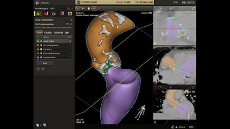
Automatic segmentation saves time

Once you load a pre-operative DICOM compliant CT dataset and a 3D volume is rendered, HeartNavigator automatically segments tissue, anatomical structures, landmarks, calcium, and planes of the heart for TAVR/TAVI. To facilitate mitral valve replacement, left atrial appendage closure (LAAC) and other procedures, HeartNavigator automatically segments the entire heart.

Automatic segmentation saves time

Automatic segmentation saves time
Once you load a pre-operative DICOM compliant CT dataset and a 3D volume is rendered, HeartNavigator automatically segments tissue, anatomical structures, landmarks, calcium, and planes of the heart for TAVR/TAVI. To facilitate mitral valve replacement, left atrial appendage closure (LAAC) and other procedures, HeartNavigator automatically segments the entire heart.

Automatic segmentation saves time

Once you load a pre-operative DICOM compliant CT dataset and a 3D volume is rendered, HeartNavigator automatically segments tissue, anatomical structures, landmarks, calcium, and planes of the heart for TAVR/TAVI. To facilitate mitral valve replacement, left atrial appendage closure (LAAC) and other procedures, HeartNavigator automatically segments the entire heart.
Click here for more information
Automatic segmentation saves time

Automatic segmentation saves time

Once you load a pre-operative DICOM compliant CT dataset and a 3D volume is rendered, HeartNavigator automatically segments tissue, anatomical structures, landmarks, calcium, and planes of the heart for TAVR/TAVI. To facilitate mitral valve replacement, left atrial appendage closure (LAAC) and other procedures, HeartNavigator automatically segments the entire heart.
Automatic landmarks to stay on track
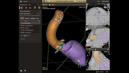
Automatic landmarks to stay on track

For additional guidance, HeartNavigator automatically places landmarks on a large variety of anatomical structures, including the ostia of the coronaries, 3 nadirs, etc. They can be manually adapted as needed.

Automatic landmarks to stay on track

Automatic landmarks to stay on track
For additional guidance, HeartNavigator automatically places landmarks on a large variety of anatomical structures, including the ostia of the coronaries, 3 nadirs, etc. They can be manually adapted as needed.

Automatic landmarks to stay on track

For additional guidance, HeartNavigator automatically places landmarks on a large variety of anatomical structures, including the ostia of the coronaries, 3 nadirs, etc. They can be manually adapted as needed.
Click here for more information
Automatic landmarks to stay on track

Automatic landmarks to stay on track

For additional guidance, HeartNavigator automatically places landmarks on a large variety of anatomical structures, including the ostia of the coronaries, 3 nadirs, etc. They can be manually adapted as needed.
Automatic measurements help avoid errors
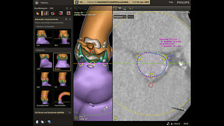

Automatic measurements help avoid errors

A single click creates area, perimeter, diameter and distance measurements of anatomical structures for TAVR or TAVI procedures. The measurements are performed within the detected anatomical planes and are shown on the displayed centerline. For all other SHD procedures, manual measurements are provided.

Automatic measurements help avoid errors

Automatic measurements help avoid errors
A single click creates area, perimeter, diameter and distance measurements of anatomical structures for TAVR or TAVI procedures. The measurements are performed within the detected anatomical planes and are shown on the displayed centerline. For all other SHD procedures, manual measurements are provided.

Automatic measurements help avoid errors

A single click creates area, perimeter, diameter and distance measurements of anatomical structures for TAVR or TAVI procedures. The measurements are performed within the detected anatomical planes and are shown on the displayed centerline. For all other SHD procedures, manual measurements are provided.
Click here for more information
Automatic measurements help avoid errors

Automatic measurements help avoid errors

A single click creates area, perimeter, diameter and distance measurements of anatomical structures for TAVR or TAVI procedures. The measurements are performed within the detected anatomical planes and are shown on the displayed centerline. For all other SHD procedures, manual measurements are provided.
Calcification visualization to avoid potential complications
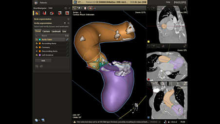

Calcification visualization to avoid potential complications

The software enhances insight into the distribution of calcifications in the ascending aorta, aortic valve annulus, and left ventricle. By determining the severity and location of calcification, you can avoid potential complications during procedures.

Calcification visualization to avoid potential complications

Calcification visualization to avoid potential complications
The software enhances insight into the distribution of calcifications in the ascending aorta, aortic valve annulus, and left ventricle. By determining the severity and location of calcification, you can avoid potential complications during procedures.

Calcification visualization to avoid potential complications

The software enhances insight into the distribution of calcifications in the ascending aorta, aortic valve annulus, and left ventricle. By determining the severity and location of calcification, you can avoid potential complications during procedures.
Click here for more information
Calcification visualization to avoid potential complications

Calcification visualization to avoid potential complications

The software enhances insight into the distribution of calcifications in the ascending aorta, aortic valve annulus, and left ventricle. By determining the severity and location of calcification, you can avoid potential complications during procedures.
Automatic view planning aids positioning
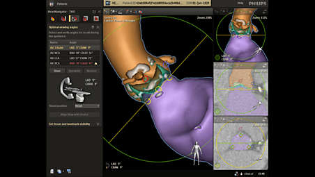

Automatic view planning aids positioning

HeartNavigator automatically determines the most optimal projection angles to use during the procedure. This can avoid the need to acquire multiple aortagrams. Projections can be recalled tableside for further savings.

Automatic view planning aids positioning

Automatic view planning aids positioning
HeartNavigator automatically determines the most optimal projection angles to use during the procedure. This can avoid the need to acquire multiple aortagrams. Projections can be recalled tableside for further savings.

Automatic view planning aids positioning

HeartNavigator automatically determines the most optimal projection angles to use during the procedure. This can avoid the need to acquire multiple aortagrams. Projections can be recalled tableside for further savings.
Click here for more information
Automatic view planning aids positioning

Automatic view planning aids positioning

HeartNavigator automatically determines the most optimal projection angles to use during the procedure. This can avoid the need to acquire multiple aortagrams. Projections can be recalled tableside for further savings.
Enhanced device selection to check correct fit
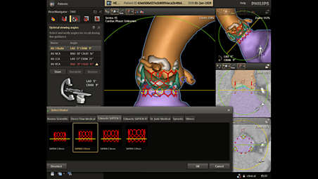

Enhanced device selection to check correct fit

It is critical in TAVR/TAVI to select a properly sized aortic valve repair device. HeartNavigator lets you visualize the 3D virtual device templates of many of the latest TAVR/TAVI devices, modeled in cooperation with leading device manufacturers.

Enhanced device selection to check correct fit

Enhanced device selection to check correct fit
It is critical in TAVR/TAVI to select a properly sized aortic valve repair device. HeartNavigator lets you visualize the 3D virtual device templates of many of the latest TAVR/TAVI devices, modeled in cooperation with leading device manufacturers.

Enhanced device selection to check correct fit

It is critical in TAVR/TAVI to select a properly sized aortic valve repair device. HeartNavigator lets you visualize the 3D virtual device templates of many of the latest TAVR/TAVI devices, modeled in cooperation with leading device manufacturers.
Click here for more information
Enhanced device selection to check correct fit

Enhanced device selection to check correct fit

It is critical in TAVR/TAVI to select a properly sized aortic valve repair device. HeartNavigator lets you visualize the 3D virtual device templates of many of the latest TAVR/TAVI devices, modeled in cooperation with leading device manufacturers.
Integrated live image guidance supports precise navigation
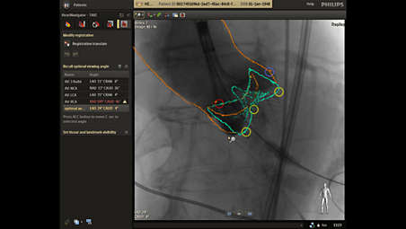

Integrated live image guidance supports precise navigation

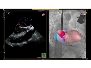
You can get real-time feedback to guide navigation through the vasculature. The 3D CT volume rendering of the ascending aorta can be matched with live fluoro to show the exact position of catheters and devices in relation to the reference image. The 3D CT rendering moves in sync with system movements during the procedure.

Integrated live image guidance supports precise navigation

Integrated live image guidance supports precise navigation
You can get real-time feedback to guide navigation through the vasculature. The 3D CT volume rendering of the ascending aorta can be matched with live fluoro to show the exact position of catheters and devices in relation to the reference image. The 3D CT rendering moves in sync with system movements during the procedure.

Integrated live image guidance supports precise navigation

You can get real-time feedback to guide navigation through the vasculature. The 3D CT volume rendering of the ascending aorta can be matched with live fluoro to show the exact position of catheters and devices in relation to the reference image. The 3D CT rendering moves in sync with system movements during the procedure.
Click here for more information
Integrated live image guidance supports precise navigation

Integrated live image guidance supports precise navigation

You can get real-time feedback to guide navigation through the vasculature. The 3D CT volume rendering of the ascending aorta can be matched with live fluoro to show the exact position of catheters and devices in relation to the reference image. The 3D CT rendering moves in sync with system movements during the procedure.
  • Automatic segmentation saves time
  • Automatic landmarks to stay on track
  • Automatic measurements help avoid errors
  • Calcification visualization to avoid potential complications
See all features
Automatic segmentation saves time

Automatic segmentation saves time

Once you load a pre-operative DICOM compliant CT dataset and a 3D volume is rendered, HeartNavigator automatically segments tissue, anatomical structures, landmarks, calcium, and planes of the heart for TAVR/TAVI. To facilitate mitral valve replacement, left atrial appendage closure (LAAC) and other procedures, HeartNavigator automatically segments the entire heart.

Automatic segmentation saves time

Automatic segmentation saves time
Once you load a pre-operative DICOM compliant CT dataset and a 3D volume is rendered, HeartNavigator automatically segments tissue, anatomical structures, landmarks, calcium, and planes of the heart for TAVR/TAVI. To facilitate mitral valve replacement, left atrial appendage closure (LAAC) and other procedures, HeartNavigator automatically segments the entire heart.

Automatic segmentation saves time

Once you load a pre-operative DICOM compliant CT dataset and a 3D volume is rendered, HeartNavigator automatically segments tissue, anatomical structures, landmarks, calcium, and planes of the heart for TAVR/TAVI. To facilitate mitral valve replacement, left atrial appendage closure (LAAC) and other procedures, HeartNavigator automatically segments the entire heart.
Click here for more information
Automatic segmentation saves time

Automatic segmentation saves time

Once you load a pre-operative DICOM compliant CT dataset and a 3D volume is rendered, HeartNavigator automatically segments tissue, anatomical structures, landmarks, calcium, and planes of the heart for TAVR/TAVI. To facilitate mitral valve replacement, left atrial appendage closure (LAAC) and other procedures, HeartNavigator automatically segments the entire heart.
Automatic landmarks to stay on track

Automatic landmarks to stay on track

For additional guidance, HeartNavigator automatically places landmarks on a large variety of anatomical structures, including the ostia of the coronaries, 3 nadirs, etc. They can be manually adapted as needed.

Automatic landmarks to stay on track

Automatic landmarks to stay on track
For additional guidance, HeartNavigator automatically places landmarks on a large variety of anatomical structures, including the ostia of the coronaries, 3 nadirs, etc. They can be manually adapted as needed.

Automatic landmarks to stay on track

For additional guidance, HeartNavigator automatically places landmarks on a large variety of anatomical structures, including the ostia of the coronaries, 3 nadirs, etc. They can be manually adapted as needed.
Click here for more information
Automatic landmarks to stay on track

Automatic landmarks to stay on track

For additional guidance, HeartNavigator automatically places landmarks on a large variety of anatomical structures, including the ostia of the coronaries, 3 nadirs, etc. They can be manually adapted as needed.
Automatic measurements help avoid errors

Automatic measurements help avoid errors

A single click creates area, perimeter, diameter and distance measurements of anatomical structures for TAVR or TAVI procedures. The measurements are performed within the detected anatomical planes and are shown on the displayed centerline. For all other SHD procedures, manual measurements are provided.

Automatic measurements help avoid errors

Automatic measurements help avoid errors
A single click creates area, perimeter, diameter and distance measurements of anatomical structures for TAVR or TAVI procedures. The measurements are performed within the detected anatomical planes and are shown on the displayed centerline. For all other SHD procedures, manual measurements are provided.

Automatic measurements help avoid errors

A single click creates area, perimeter, diameter and distance measurements of anatomical structures for TAVR or TAVI procedures. The measurements are performed within the detected anatomical planes and are shown on the displayed centerline. For all other SHD procedures, manual measurements are provided.
Click here for more information
Automatic measurements help avoid errors

Automatic measurements help avoid errors

A single click creates area, perimeter, diameter and distance measurements of anatomical structures for TAVR or TAVI procedures. The measurements are performed within the detected anatomical planes and are shown on the displayed centerline. For all other SHD procedures, manual measurements are provided.
Calcification visualization to avoid potential complications

Calcification visualization to avoid potential complications

The software enhances insight into the distribution of calcifications in the ascending aorta, aortic valve annulus, and left ventricle. By determining the severity and location of calcification, you can avoid potential complications during procedures.

Calcification visualization to avoid potential complications

Calcification visualization to avoid potential complications
The software enhances insight into the distribution of calcifications in the ascending aorta, aortic valve annulus, and left ventricle. By determining the severity and location of calcification, you can avoid potential complications during procedures.

Calcification visualization to avoid potential complications

The software enhances insight into the distribution of calcifications in the ascending aorta, aortic valve annulus, and left ventricle. By determining the severity and location of calcification, you can avoid potential complications during procedures.
Click here for more information
Calcification visualization to avoid potential complications

Calcification visualization to avoid potential complications

The software enhances insight into the distribution of calcifications in the ascending aorta, aortic valve annulus, and left ventricle. By determining the severity and location of calcification, you can avoid potential complications during procedures.
Automatic view planning aids positioning

Automatic view planning aids positioning

HeartNavigator automatically determines the most optimal projection angles to use during the procedure. This can avoid the need to acquire multiple aortagrams. Projections can be recalled tableside for further savings.

Automatic view planning aids positioning

Automatic view planning aids positioning
HeartNavigator automatically determines the most optimal projection angles to use during the procedure. This can avoid the need to acquire multiple aortagrams. Projections can be recalled tableside for further savings.

Automatic view planning aids positioning

HeartNavigator automatically determines the most optimal projection angles to use during the procedure. This can avoid the need to acquire multiple aortagrams. Projections can be recalled tableside for further savings.
Click here for more information
Automatic view planning aids positioning

Automatic view planning aids positioning

HeartNavigator automatically determines the most optimal projection angles to use during the procedure. This can avoid the need to acquire multiple aortagrams. Projections can be recalled tableside for further savings.
Enhanced device selection to check correct fit

Enhanced device selection to check correct fit

It is critical in TAVR/TAVI to select a properly sized aortic valve repair device. HeartNavigator lets you visualize the 3D virtual device templates of many of the latest TAVR/TAVI devices, modeled in cooperation with leading device manufacturers.

Enhanced device selection to check correct fit

Enhanced device selection to check correct fit
It is critical in TAVR/TAVI to select a properly sized aortic valve repair device. HeartNavigator lets you visualize the 3D virtual device templates of many of the latest TAVR/TAVI devices, modeled in cooperation with leading device manufacturers.

Enhanced device selection to check correct fit

It is critical in TAVR/TAVI to select a properly sized aortic valve repair device. HeartNavigator lets you visualize the 3D virtual device templates of many of the latest TAVR/TAVI devices, modeled in cooperation with leading device manufacturers.
Click here for more information
Enhanced device selection to check correct fit

Enhanced device selection to check correct fit

It is critical in TAVR/TAVI to select a properly sized aortic valve repair device. HeartNavigator lets you visualize the 3D virtual device templates of many of the latest TAVR/TAVI devices, modeled in cooperation with leading device manufacturers.
Integrated live image guidance supports precise navigation

Integrated live image guidance supports precise navigation

You can get real-time feedback to guide navigation through the vasculature. The 3D CT volume rendering of the ascending aorta can be matched with live fluoro to show the exact position of catheters and devices in relation to the reference image. The 3D CT rendering moves in sync with system movements during the procedure.

Integrated live image guidance supports precise navigation

Integrated live image guidance supports precise navigation
You can get real-time feedback to guide navigation through the vasculature. The 3D CT volume rendering of the ascending aorta can be matched with live fluoro to show the exact position of catheters and devices in relation to the reference image. The 3D CT rendering moves in sync with system movements during the procedure.

Integrated live image guidance supports precise navigation

You can get real-time feedback to guide navigation through the vasculature. The 3D CT volume rendering of the ascending aorta can be matched with live fluoro to show the exact position of catheters and devices in relation to the reference image. The 3D CT rendering moves in sync with system movements during the procedure.
Click here for more information
Integrated live image guidance supports precise navigation

Integrated live image guidance supports precise navigation

You can get real-time feedback to guide navigation through the vasculature. The 3D CT volume rendering of the ascending aorta can be matched with live fluoro to show the exact position of catheters and devices in relation to the reference image. The 3D CT rendering moves in sync with system movements during the procedure.

You are about to visit a Philips global content page

Continue

You are about to visit a Philips global content page

Continue

Site-ul nostru poate fi vizualizat cel mai bine cu cea mai recentă versiune de Microsoft Edge, Google Chrome sau Firefox.

Accesați un website-ul Philips Healthcare România
Please select the checkbox

Conținutul paginii următoare conține informații destinate exclusiv profesioniștilor din domeniul sănătății, întrucât se referă la produse care se încadrează în categoria dispozitivelor medicale care necesită utilizarea sau intervenția profesioniștilor din sectorul medical.

Ești o persoană care întrunește criteriile de mai sus?